Home
/ Back Of Head Skull Anatomy - Posterior and Lateral views of the Skull - Anatomy | Kenhub - Anatomical head model, anatomical human anatomical half head and face anatomy medical brain neck median section study model.
Back Of Head Skull Anatomy - Posterior and Lateral views of the Skull - Anatomy | Kenhub - Anatomical head model, anatomical human anatomical half head and face anatomy medical brain neck median section study model.
Back Of Head Skull Anatomy - Posterior and Lateral views of the Skull - Anatomy | Kenhub - Anatomical head model, anatomical human anatomical half head and face anatomy medical brain neck median section study model.. The skull is the bony skeleton of the head. The skull encases and protects the brain as well as the special sense organs of vision, hearing, balance, taste and smell. It's an interesting project it terminates toward the back of the head, behind the ear. It offers protection to the brain, eye balls, inner ears, and nasal passages. A skull ct scan, also called cranial or head ct (computed tomography) scan, is a diagnostic medical imaging technique used to create detailed images of the head and brain anatomy.
This anatomic region is complex and poses surgical challenges for otolaryngologists and neurosurgeons alike. Bone that forms the forehead. Skull, skeletal framework of the head of vertebrates, composed of bones or cartilage, which form a unit that protects the brain and some sense organs. The cranium and the mandible. Skull reshaping is done on any of the structures that lie above the face.
Human Skull Anatomy Illustration 1866 Antique Textbook ... from cdn11.bigcommerce.com It is the collection of 22 bones, settled by intramembranous ossification, that is joined together by sutures identified as the fibrous joint. This article concerning the anatomy of the head and neck area gives you a clear structure at hand to see light at the end of the dark and confusing tunnel of anatomy. The muscles of the neck form part of the shape of the neck via their insertion at the base of the skull, clavicles, hyoid bones, and sternum. Anatomical head model, anatomical human anatomical half head and face anatomy medical brain neck median section study model. In the adult, the skull consists of 22 individual bones, 21 of which are immobile and united into a single unit. In order to be light, the skull is made up by flat and irregular bones, and has hollow spaces called the sinuses. The skull begins to form prior to week 12 of embryogenesis. It offers protection to the brain, eye balls, inner ears, and nasal passages.
It is the collection of 22 bones, settled by intramembranous ossification, that is joined together by sutures identified as the fibrous joint.
The skull is the skeleton of the head. The skull also supports tendinous muscle attachments and allows neurovascular passage between intracranial and extracranial anatomy. The combination of skull and surface anatomy in this study is quite macabre. Learn about anatomy skull with free interactive flashcards. The cranium and mandible was exported from ct data. These individual plates of bone fuse together after. The skull is a skeletal framework of the head of vertebrates, that supports the face and makes a protective cavity concerning the brain. Continue scrolling to read more below. A human skull is almost full sized at birth. Note also the quite acute angle formed by. The cranium (skull) is the skeletal structure of the head that supports the face and protects the brain. In order to be light, the skull is made up by flat and irregular bones, and has hollow spaces called the sinuses. We discuss the anatomy, function and pathologies of the skull bones.
This means that the skull can flex and deform during birth, making it easier to deliver a baby through the narrow birth canal. The cranium and mandible was exported from ct data. Note also the quite acute angle formed by. Foramina inside the body of humans and other animals. Pain in the back of your head at the base of your skull can cause your head to hurt with dull, nagging persistent pains.
Lowpoly Human Skull 3D Model from d1a9v60rjx2a4v.cloudfront.net This is a model of the human (homo sapiens) skull. The cranium and mandible was exported from ct data. We discuss the anatomy, function and pathologies of the skull bones. It is comprised of many bones, formed by intramembranous ossification, which are joined together by sutures (fibrous joints). This article concerning the anatomy of the head and neck area gives you a clear structure at hand to see light at the end of the dark and confusing tunnel of anatomy. Skull, skeletal framework of the head of vertebrates, composed of bones or cartilage, which form a unit that protects the brain and some sense organs. The skull is a bone structure that forms the head in vertebrates. Note also the quite acute angle formed by.
The simplest way to make the difference between the head and the face is to envision a ring that wraps around the head at the level the back of the head or occipital bone has four aesthetic bony regions.
However the eight bones that make up the cranium are not yet fused together. Learn more about the anatomy and function of the skull in humans and other vertebrates. From an anatomical perspective, the skull is divided into two parts: The cranium and mandible was exported from ct data. In order to be light, the skull is made up by flat and irregular bones, and has hollow spaces called the sinuses. If you have a trapped nerve in your cervical spine, you may experience sharp jabbing pains that radiate to your temples or behind your eye. Continue scrolling to read more below. It was then cleaned, adapted and polypainted this model is part of a comparison with the skull of a human. Please feel free to download and print. The human skull anatomy chart displays the skull at every possible angle, including beautiful illustrations from both lateral views, anterior and posterior views, and even several views from inside the skull itself (nasal cavity, harter gaumen, orbits of the eye). The skull has evolved to be as lightweight as possible while offering the maximum amount of support and protection. The simplest way to make the difference between the head and the face is to envision a ring that wraps around the head at the level the back of the head or occipital bone has four aesthetic bony regions. The cranium (skull) is the skeletal structure of the head that supports the face and protects the brain.
We discuss the anatomy, function and pathologies of the skull bones. It is comprised of many bones, formed by intramembranous ossification, which are joined together by sutures (fibrous joints). Skull reshaping is done on any of the structures that lie above the face. Due to its complex development and associated important structures, understanding skull anatomy holds abundant clinical and surgical significance. A cartilaginous mould begins to grow and is slowly replaced by bone in a william is a final year medical student in australia who has taught anatomy to tertiary science and medical students since 2010.
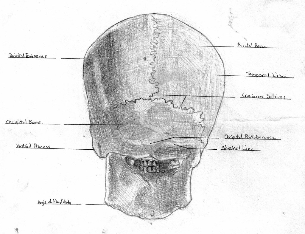
Anatomy: Skull back | Greg Allen | Flickr from c1.staticflickr.com Anatomy of the skull and bones of cranium on medical illustrations. The cranium and mandible was exported from ct data. Due to its complex development and associated important structures, understanding skull anatomy holds abundant clinical and surgical significance. In the adult, the skull consists of 22 individual bones, 21 of which are immobile and united into a single unit. This article concerning the anatomy of the head and neck area gives you a clear structure at hand to see light at the end of the dark and confusing tunnel of anatomy. Skull, skeletal framework of the head of vertebrates, composed of bones or cartilage, which form a unit that protects the brain and some sense organs. If you have a trapped nerve in your cervical spine, you may experience sharp jabbing pains that radiate to your temples or behind your eye. Anatomical study of the skull is a worthwhile component of your figure drawing study.
Cranial cavity , cranial sutures.
Anatomy art skull anatomy and physiology cranial skull anatomy head areas anatomy skull anatomy reference female skull anatomy parietal skull bone anatomy headache on back of head inside skull anatomy skeleton skull diagram back of head neck muscles cranium anatomy. This anatomic region is complex and poses surgical challenges for otolaryngologists and neurosurgeons alike. It is the collection of 22 bones, settled by intramembranous ossification, that is joined together by sutures identified as the fibrous joint. The skull encases and protects the brain as well as the special sense organs of vision, hearing, balance, taste and smell. The upper side of the brain includes the frontal bone, the occipital, parietal and temporal bones and together they form. Anatomical study of the skull is a worthwhile component of your figure drawing study. In the adult, the skull consists of 22 individual bones, 21 of which are immobile and united into a single unit. The skull is a bone structure that forms the head in vertebrates. These joints fuse together in adulthood. We discuss the anatomy, function and pathologies of the skull bones. The sagittal suture is the line where the right and left parietal bone are in contact. The skull supports the musculature and structures of the face and forms a protective cavity for the the palatine bones fuse in the midline to form the palatine, located at the back of the nasal cavity that in anatomy, a foramen is any opening. Bone that forms the forehead.
A human skull is almost full sized at birth back of skull anatomy. This means that the skull can flex and deform during birth, making it easier to deliver a baby through the narrow birth canal.

